Art and science combine in the “Body Parts” exhibit
UW-Eau Claire students and local professional artists are taking the practice of studying the human body and transforming it into a new medium, art
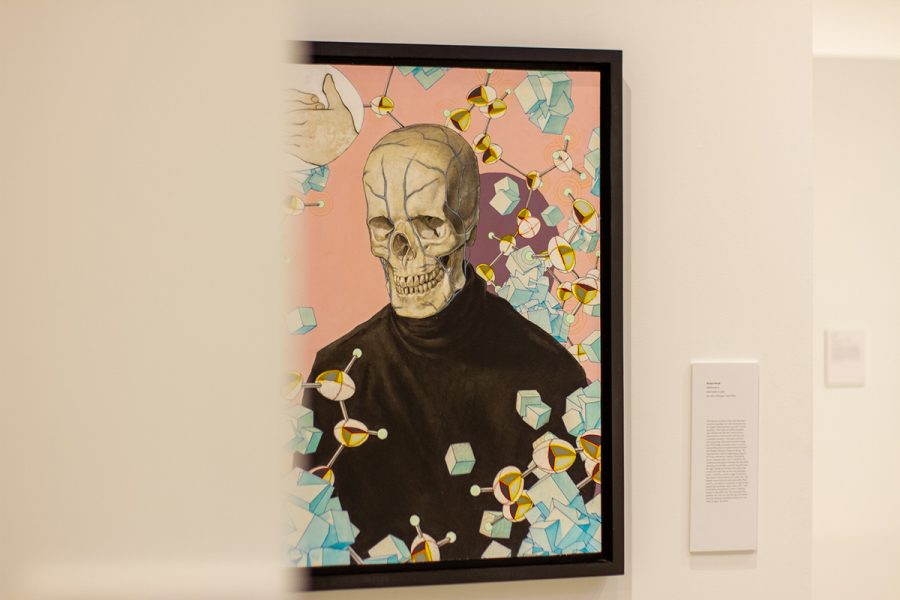
Photo by McKenna Dirks
A piece of art on display for the “bones” part of the Body Parts exhibit.
In partnership with the University of Wisconsin-Eau Claire’s Institute for Health Sciences, UW-Eau Claire’s Foster Gallery is showcasing “Body Parts,” an exhibition where the university’s artists explore six of the human body’s organs and depict biology through artistry.
Free for Blugolds and the public, the “Body Parts” exhibit provides a unique perspective on the human form. What is typically untouched by art and left to the realms of science is being transfigured into the metaphysical and learned through creation.
The exhibition owes its actualization to its curators: Mel Kantor, an endowed chair of the Institute of Health and Sciences, and Jill Olm, an associate professor of art and design.
With the help of the art and design and biology faculty as well as Eau Claire radiologist Alicia Arnold, students and members of the Eau Claire community can see their bodies in a new way.
Influenced by modern medical devices and tools like X-rays, medical images and photomicrographs, artists and students were able to take a microscopic look into what makes the human body work and reinterpret the anatomy in their own way.
For Jack Degan, a third-year graphic communications student, he said the process of using scientific reference for artistic design and how the craft is not as easy as it might look.
“I learned that if you don’t reference it exactly it won’t look how you want it,” Degan said. “The brain and the heart are very detailed so if I didn’t add a certain crease or fold it would be easy to identify.”
Besides student artwork, visitors can expect the opportunity to see pieces created by artists who don’t attend UW-Eau Claire as well.
One such artist is Kindra Crick, a multimedia artist whose mixed-media interpretations give visual expression to the process of scientific discovery. Crick uses her degree in Molecular Biology from Princeton and her Certificate in Painting from the School of Art Institute in Chicago to combine two opposite, complex concepts. Crick incorporates drawing, diagrams, maps and microscopic imagery to create her art.
While discussing how Leonardo DaVinci’s anatomical drawings inspired her art, she points out the equal importance of both describing and depicting.
“Not only did he write about the descriptions of the anatomy but he knew that it was important to depict the anatomy because that somehow penetrated our understanding,” Crick said. “That idea of observation is really important.”
Crick’s work has earned her numerous features in magazines like Huffington Post, The daVinci Pursuit and PBS Newshour. She has also had work exhibited in a variety of shows in both the US and the UK, such as the New York Hall of Science, MDI Biological Laboratory and Cambridge, England’s LMB permanent collection.
The “Body Parts” art exhibition is set to last from Feb. 4 to March 9 and is open from 10:00 a.m. to 4:30 p.m. seven days a week. The Gallery has masking requirements in place so make sure to mask up before taking a trip through the human body’s organs.
Hinrichs can be reached at hinricha0521.uwec.edu

Allison Hinrichs is a third-year journalism and multimedia communication student. This is her fourth semester at The Spectator. Allison loves being outdoors and anything that gives her an adrenaline rush. She loves hiking, rock climbing, snowboarding and photography.

McKenna Dirks is a fourth-year journalism student and this is her seventh semester on The Spectator staff. She thrives under chaotic environments, loves plants and often gives off "granola girl" vibes with her Blundstone boots.


Angie • Oct 29, 2022 at 3:38 am
I’m very interested in this event coz I’m obsessed with skulls